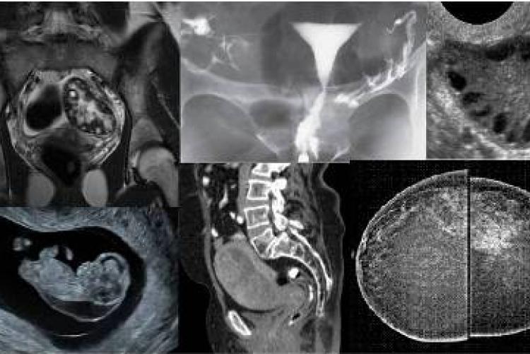
Women's Imaging encompasses the full range of imaging services for breast, gynecologic and obstetric Imaging. Breast Imaging is a subspecialty of radiology which focuses on both screening and diagnostic imaging procedures that include mammography, breast ultrasound and breast MRI. Breast biopsies are also performed as part of breast cancer diagnosis.
Related resident dissertations include the following:
Current research projects:
None
MMed Thesis:
Breast Imaging
-
Kuppuswamy Naaila Balaraman (2021). A comparison between digital breast tomosynthesis and ultrasound in the characterization of mammographic breast lesions using histopathology as the gold standard at KNH.
-
Ndlovu Sijabule Swodziwa (2021). The added value of sonoelastography in evaluating breast masses detected in mammography.
-
Sedheva Manpreet Kaur (2021). Prevalence of mammographic calcifications in KNH.
-
Mose Linda (2020). The role of digital breast tomosynthesis as an adjunct to digital mammography in the characterization of breast lesions at KNH.
-
Ndaiga, P. W. (2015). Evaluation of diagnostic accuracy of ultrasound elastography in differentiating benign and malignant solid breast masses.
-
Singh, L. S. (2013).Validity of BIRAD system in mammography reporting.
-
Kebuka, C. W. (2012).Correlation of breast Malignancies on MRI and histopathology.
-
Muruka, J. (2012). Determination of radiation dose in bilateral view mammography.
-
Kagia, J.N. (2011).Correlation between ultrasonography and histopathology in Breast Abnormalities at Kenyatta National Hospital.
-
Mutunga, C.M.S. (2004). The value of ultrasonography as an adjunct to mammography in evaluating breast masses.
-
Mwangi, J.K. (1996). Usefulness of mammography in the investigation of symptomatic patients under 30 years of age at Kenyatta National Hospital.
-
Vinayak,S. (1988). Mammography.
Obstetric and Gynaecological Imaging
-
Kitili Martha Ndanu (2021). Correlation of uterine artery Doppler velocimetry with laboratory findings in patients with severe pre-eclampsia at KNH.
-
Mustansir Nanabhai B. (2021). Determination of Local reference ranges of uterine and umbilical artery Doppler indices: A Kenyatta National Hospital Experience
-
Mukaindo Caroline (2020). Evaluating the accuracy of transcerebellar diameter in estimation of gestation age in second and third trimester at KNH
-
Mosomi Abigael (2020). Ultrasonographic Evaluation of Placental Thickness in normal singleton pregnancies for estimation of gestational age in Kenyata National Hospital.
-
Mwagiru Josephine W. (2020). Ultrasonographic Assessment of Fetal epiphyseal ossification centres for estimation of gestational age in the third trimester.
-
Achan, M.A. (2016). Hysterosalpingography findings in the infertile patient in Kenyatta National Hospital.
-
Githinji, I.N. (2014). Sonographic findings in patients with first trimester bleeding.
-
Parmar, L. (2013). Determination of fetal outcome in patients in 3rd trimester with hypertensive disorders of pregnancy.
-
Swaleh, M.M. (2013). Sonohysterography findings in abnormal uterine bleeding in Kenyatta National Hospital.
-
Mamai, C.A. (2009). The role of MRI in the evaluation of gynecological disorders.
-
Nshizirungu, J.J. (2009). A correlation of ultrasound and surgical findings in suspected ectopic pregnancy at Kenyatta National Hospital.
-
Gikonyo, J.M. (2006). Correlation between ultrasonography and histopathological findings of gynaecological pelvic masses at Kenyatta National Hospital.
-
Mundia, J.G. (2004). Intrapartum sonogrpahic foetal weight estimation/ the actual birth weight at Pumwani Maternity Hospital.
-
Nguku, S.W. (2003). The biophysical profile scores and resistive indices of the umbilical artery as seen on patients with pregnancy induced hypertension at two Nairobi hospitals.
-
Manduku, V. (2003). A comparison of hysterosalpingography and laparoscopy findings in the diagnosis of tubal factors in infertile women.
-
Kimani, N.M. (2000).The pattern of female pelvic disease as shown by ultrasound.
-
Butt, W. (1999). A correlative study of the various gestational age parameters of our local population on ultrasound.
-
Kimaru, J. (1995). Common Technical Errors in Hysterosalpingography (HSG).
-
Wanga, H.O.J. (1995). Ultrasonographic foetal age assessment versus menstrual age in the second and third trimester.
-
Ntarangwi, F.N. (1993). Comparison of ultrasonography with clinical examination under anaesthesia in the staging of carcinoma of the cervix at KNH.
-
Ndushabandi, D. (1989). The role of ultrasonography in management of gynaecologic pelvic masses at KNH. A one year prospective study.
-
Wambugu, M.N. (1982). The “Ten Day Rule” and its implementation at the Kenyatta National Hospital.
Faculty Lead: